
CERC Dr. Yanwen Zhang awarded the Ontario Research Fund - Small Infrastructure Fund (ORF-SIF)
Congratulations to Canada Excellence Research Chair (CERC) Dr. Yanwen Zhang for receiving the Ontario Research Fund – Small Infrastructure Fund (ORF-SIF)!
Congratulations to Canada Excellence Research Chair (CERC) Dr. Yanwen Zhang for receiving the Ontario Research Fund – Small Infrastructure Fund (ORF-SIF)!
Start Date
Tuesday April 15, 2025End Date
Thursday April 17, 2025Time
9:00 am - 5:00 pmLocation
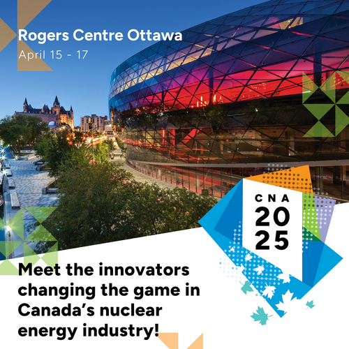
Rogers Centre OttawaCanada is increasing its momentum in the development of nuclear energy. What additional initiatives must Canada undertake to actively support nuclear projects, establish regulatory frameworks, and encourage public-private partnerships to enhance Canada’s role in clean energy production? Queen's nuclear materials group is ready to join the conversation at #CNA2025, being held April 15 to 17. We can’t wait to meet you there!
Student program application deadline: January 31, 2025
For more information and to register, visit the CNA website.
Razieh Larki (PhD student) and Richard Meng (Master’s student), from the Queen’s nuclear groups, won top honors at the UNENE Annual R&D Workshop 2024 Student Symposium, which took place in Toronto, Ontario, on December 9-10, 2024.
The Nuclear Materials Group hosted Prof. Harry J. Whitlow from Uppsala University (Sweden) on October 18 at McLaughlin Hall, as part of the Mechanical and Materials Engineering's Distinguished Speakers Series. Dr.
Date
Saturday October 19, 2024Location
ZoomJoin this exciting and insightful webinar on “Nuclear Materials” hosted by the Department of Design, IIT Roorkee in association with the Nuclear Materials Group at Queen's University, Canada, under the banner of the International Symposium IDEAS-2024.
This webinar is designed for undergraduate, postgraduate, and PhD students, as well as faculty members interested in exploring the future of nuclear energy and materials science.
Date: Saturday, October 19, 2024
Time: 5:30 PM – 7:30 PM IST | 8:00 AM - 10:00 AM EST
Platform: Zoom (Link will be shared after registration)
Registration:
No additional registration fee is required to be paid, but filling up of the registration form is mandatory.
Please register for the event using the following link: Webinar Registration
Agenda:
Introduction to the Nuclear Materials Group and RMTL
Micromechanisms of deformation in metals, ceramics, and composites
- Prof. Mark Daymond
Irradiation effects on the performance of structural materials and characterization
- Prof. Levente Balogh
Defects and defect engineering in complex materials: Experiments and modeling
- Profs. Yanwen Zhang and Laurent Béland
Microstructure and properties of nuclear materials and composite coatings
- Prof. Zhongwen Yao
Corrosion and high temperature oxidation of metals and alloys used in nuclear power plants
- Prof. Suraj Persaud
Get to Know Your Speakers:
The Nuclear Materials Group is committed to advancing nuclear energy as a much-needed part of Canada's future low carbon and sustainable energy plan. The group is internationally competitive with very strong industrial connections.
Six faculty members form a core team.
Prof. Mark R. Daymond leads the Nuclear Materials Group and is an expert in radiation-induced deformation mechanisms in nuclear structural alloys, with a focus on thermomechanical properties, slip, twinning, phase transformations, cracking, material characterization, and crystal plasticity modeling.
Prof. Levente Balogh focuses on advanced X-ray and neutron diffraction techniques to study radiation-induced damage and microstructural changes in materials.
Prof. Laurent K. Béland develops and applies computational methods to address science and engineering challenges, drawing on principles from physical chemistry, quantum mechanics, statistical physics, solid mechanics, and mathematical modeling.
Prof. Suraj Persaud specializes in corrosion and high-temperature oxidation of metals and alloys, particularly those used in nuclear power plant components.
Prof. Zhongwen Yao is an expert in microstructural characterization, focusing on radiation damage, dislocations, vacancy defects, and defect clusters in nuclear materials.
Prof. Yanwen Zhang explores equilibrium and non-equilibrium defect dynamics, ion beam modification, and radiation effects in materials, with an emphasis on tailoring the functionality and properties of complex materials.
Profs. Mark Daymond and Suraj Persaud in Mechanical and Materials Engineering, and Prof. Warren Mabee in Policy Studies have received a multi-disciplinary grant from Natural Resources Canada’s Enabling Small Modular Reactors (SMR) Program.
Date
Friday October 18, 2024Location
Room 306 McLaughlin HallDistinguished Speakers Series
MeV ion microprobes for study of radiation damage
by Dr. Harry J. Whitlow, Uppsala University, Sweden
Friday, October 18, 2024
10:30 AM - 11:30 AM
Room 306 McLaughlin Hall
MeV ion microprobes have a long history in the study of the interactions of energetic ions with materials. Regularly, μm-sized beams can be obtained. These have been widely employed in ion beam analysis e.g. Particle Induced X-Ray Emission (PIXE) imaging. Today, for some special cases super-resolutions of a few nm have been demonstrated. Ion beam modification studies with microprobes generally exploit the extremely high fluxes attainable, for spatial writing of radiation damage in different classes of materials.
This presentation discusses some applications and potential applications of MeV ion microprobes for different aspects of radiation damage studies. What is especially useful is the high fluxes (>1015 ions cm–2 s–1) of MeV ions that can be directed into a small area (~1 μm x 1 μm) leaving surrounding areas pristine. Apart from using the radiation damage as the basis of ion beam lithography aka proton beam writing, the microprobe lens system can be utilised to perform broad beam irradiation with extremely low uniform fluxes in the range 105 – 1010 ions cm–2s–1. The latter is useful for radiation biomedicine as these fluxes correspond to absorbed dose rates in the range 10 mGy s-1. This enables studies of tissue damage and the onset acute radiation syndrome (~10 mGy) and also higher absorbed doses which are important in Ion Beam Cancer Therapy (IBCT).
Space charged-particle and neutron irradiation effects in low earth orbits and long-duration lunar and interplanetary missions are a serious problem. Energetic GeV particles (such as 56Fe ) cause soft-upsets in Metal Oxide Semiconductor (MOS) devices which are detrimental to spacecraft control and data integrity. However, low energy (<200 MeV) protons are about 104 times more abundant and damage bipolar devices, such as power switches and photovoltaic panels, as well as optical coatings as those used on camera lenses.
Hydrogen is the most abundant element and as a constituent of water plays a major role in corrosion, particularly in radiation environments where both electrolysis and radiolysis take place. The ubiquitousness of hydrogen makes it hard to analyse by e.g. mass spectroscopic methods due to contamination signals. Recently, we have developed Off-axis Scanning Transmission Ion Microscopy (OA-STIM) to quantitatively map H concentrations in self-supporting thin films (such as used in electron microscopy). This was developed for measuring C, H, N and O in biological tissue sections. However, the method is quite general and can be applied to measure and map hydrogen contents of e.g. ceramics and/or metal samples.
Speaker profile:
Harry J. Whitlow (HJW) was born in London, UK in 1954 and was educated in the UK. HJW holds a BSc (hons) and DSc degrees from the University of Bath UK, a MSc from the CNAA and a DPhil from the University of Sussex. He is a docent at the Royal Institute of Technology (KTH) in Stockholm, Sweden.
HJW’s highly international career started as a postdoc at the University of Aarhus Denmark. It includes periods working at the Universities of Helsinki and Jyväskylä in Finland, Uppsala, KTH and Malmö universities in Sweden, Haute Ecole Arc Ingénierie, University of Applied Sciences of Western Switzerland and University of Louisiana at Lafayette USA and the University of Oslo.
HJW was appointed to professorships in Lund University in 2000, Jyväskylä in 2004 and Lafayette in 2016. Today, he holds adjunct professorships at Uppsala University Sweden and Kasetsart University in Thailand and is a guest researcher in Oslo University, Norway. He has supervised 13 PhD students, his scientific production approaches 300 publications. His notable research contributions cover a wide range of topics and include surface segregation during sputtering, Monte Carlo and Molecular dynamics studies of collision cascade anisotropies, Multi-dispersive ToF-E elastic recoil detection analysis, stopping measurements, Programable proximity aperture lithography, low flux irradiation with MeV ion microprobes and more recently a quantitative microprobe method for measuring ex-vivo trace element molarity is biomedical tissues.
Research Administrative Coordinator
Mechanical & Materials Engineering
Smith Engineering
MASc Student